
AI in Medicine Science Fair Project Ideas & Step-by-Step Guide
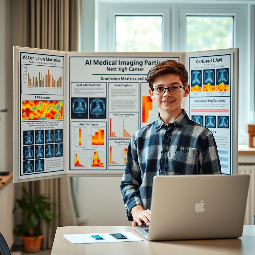
Artificial intelligence is rapidly transforming modern healthcare, from automated radiology screening to predicting patient outcomes. For middle and high school students interested in computer science and medicine, building an ai in medicine science fair exmple project provides an exceptional platform to demonstrate technical competence while addressing real-world biomedical challenges.
Science fair judges value projects that combine rigorous methodology, clear data science principles, and practical medical relevance. This guide walks you through actionable project ideas, publicly available healthcare datasets, model evaluation techniques, and competition guidelines.
Choosing a High-Impact Healthcare AI Science Fair Project
When selecting an ai in medicine science fair exmple project, it is essential to align the complexity of the machine learning task with your current programming background. Healthcare AI generally falls into three main technical domains:
- Computer Vision (Medical Imaging): Analyzing two-dimensional or three-dimensional clinical images like X-rays, retinal scans, dermatoscopic photos, or MRI scans.
- Predictive Analytics (Tabular Clinical Data): Processing structured electronic health records (EHR), vital signs, and lab parameters to forecast disease risk or patient readmission.
- Sequential & Signal Processing (Biomedical Signals): Analyzing continuous time-series data such as electrocardiograms (ECG) or electroencephalograms (EEG).
Choosing a focused, achievable hypothesis ensures you can complete training, validation, and analysis within your project timeline.
Top 4 AI in Medicine Science Fair Project Ideas
1. Skin Lesion Classification (Melanoma vs. Benign)
Detecting skin cancer early saves lives. In this computer vision project, you train a convolutional neural network (CNN) to classify images of skin moles as benign or malignant. Using transfer learning with models like ResNet or EfficientNet, students can achieve strong classification accuracy without requiring supercomputers.
2. Pneumonia Detection from Chest Radiographs
Building an ai in medicine science fair exmple project that detects pneumonia from radiology scans helps highlight how AI assists radiologists in high-volume triage. Students analyze chest X-ray images to identify pulmonary consolidations caused by bacterial or viral infections.
3. Automated Detection of Cardiac Arrhythmias
Continuous heart monitoring devices generate massive amounts of ECG data. This project uses machine learning to classify single-lead ECG wave forms into normal sinus rhythm, atrial fibrillation, or ventricular premature beats, helping identify silent heart conditions.
4. Predicting Heart Disease Risk using Clinical Variables
Using patient risk factors such as blood pressure, cholesterol levels, resting heart rate, and age, you can build a classification model to predict the presence of coronary artery disease. This project focuses on tabular data processing and model interpretability.
Trusted Public Medical Datasets for Student Researchers
Acquiring clean, ethical, and anonymized medical data is critical for a valid scientific investigation. Never attempt to collect raw medical patient data independently. Instead, leverage these reputable, open-access databases:
- NIH ChestX-ray14: Maintained by the National Institutes of Health, containing over 100,000 anonymized chest X-ray images with chest pathology labels.
- PhysioNet: A rich repository of open-access medical research data hosted by the MIT Laboratory for Computational Physiology. You can access time-series signals like the MIT-BIH Arrhythmia Database directly on PhysioNet.
- ISIC Archive: The International Skin Imaging Collaboration offers thousands of high-resolution, expert-annotated dermatoscopic skin lesion images available on the ISIC Archive.
- Kaggle Datasets: Excellent for beginners, offering curated CSV files and image folders for diabetic retinopathy, heart disease, and brain tumor MRI classification on Kaggle.
Step-by-Step Execution: From Raw Data to Model Evaluation
A structured workflow ensures your science fair experiment remains scientifically sound and repeatable.
Step 1: Define Your Research Question
Formulate a clear scientific hypothesis. For example: "Can a fine-tuned MobileNetV2 architecture achieve over 90% sensitivity in distinguishing viral pneumonia from bacterial pneumonia on chest X-rays while keeping false negatives below 5%?"
Step 2: Data Preprocessing and Cleaning
A critical phase in any ai in medicine science fair exmple project is data cleaning and preprocessing. Raw clinical datasets often suffer from missing values, class imbalances, or inconsistent image dimensions.
- For Images: Resize all images to standard dimensions (e.g., 224×224 pixels), normalize pixel intensity values between 0 and 1, and apply data augmentation (rotation, slight zooms) to prevent overfitting.
- For Tabular Data: Handle missing entries using median imputation, encode categorical variables into numerical format, and scale features using standardization.
Step 3: Model Architecture Selection & Training
Split your dataset into three distinct subsets:
- Training Set (70%): Used by the algorithm to learn features.
- Validation Set (15%): Used during training to tune hyperparameters and prevent overfitting.
- Test Set (15%): Held out until final testing to evaluate true generalization performance.
Step 4: Rigorous Evaluation
Never evaluate medical models on training data. Report overall test performance on unseen samples to prove model reliability.
Understanding Medical AI Metrics Beyond Accuracy
In standard computer science projects, simple accuracy (percentage of correct predictions) is commonly reported. In medical diagnostics, relying solely on accuracy can be dangerous due to class imbalance.
For instance, if 95% of patients in a dataset are healthy and 5% have a rare disease, a model that simply predicts every patient is healthy achieves 95% accuracy—yet fails to identify a single sick individual.
Science fair judges will expect you to calculate and explain these clinical metrics:
- Sensitivity (Recall): The proportion of actual positive cases (sick patients) correctly identified by the model. High sensitivity minimizes missed diagnoses (false negatives).
- Specificity: The proportion of actual negative cases (healthy patients) correctly identified. High specificity minimizes unnecessary treatments (false positives).
- Precision: The probability that a positive prediction by the model is accurate.
- F1-Score: The harmonic mean of precision and sensitivity, balancing both metrics.
- AUC-ROC Curve: Area Under the Receiver Operating Characteristic Curve measures the model's capacity to discriminate between diseased and non-diseased states across various decision thresholds.
Ethical Considerations and Science Fair Competition Rules
When entering events governed by International Science and Engineering Fair (ISEF) standards, strict rules apply to projects involving human subjects or health data:
- De-Identified Public Data: Using pre-existing, fully anonymized public datasets (such as NIH or PhysioNet) does not count as human subject research and typically does not require Institutional Review Board (IRB) approval prior to experiment initiation.
- Algorithmic Bias: Assess whether your training dataset disproportionately represents specific patient demographics (e.g., skin tone representation in dermatology datasets). Address potential diagnostic bias directly on your presentation board.
- Medical Advice Disclaimer: Clearly state on your display board that your AI model is intended strictly as a research prototype or decision-support tool, not as a replacement for clinical diagnosis by licensed healthcare professionals.
How to Present Your AI Medical Project to Competition Judges
Presenting your ai in medicine science fair exmple project effectively to science fair judges requires balancing code metrics with medical relevance. Follow these presentation guidelines:
- Highlight the Clinical Need: Begin your poster display by explaining the medical problem, patient burden, and how AI can improve diagnostic speed or accuracy.
- Display Confusion Matrices: Include a visual confusion matrix showing True Positives, False Positives, True Negatives, and False Negatives.
- Explain Model Interpretability: Use visualization techniques like Grad-CAM (Gradient-weighted Class Activation Mapping) for medical images to generate heatmaps. This proves your model focuses on clinical features (like lung opacities) rather than background artifacts (like patient position tags).
- Demonstrate Live Software: If permitted, bring a laptop to showcase a working demonstration where judges can input an unseen image or clinical profile and observe real-time predictions.
Related Articles
 AI
AI
Top AI Avatar Tools for Multilingual Voiceovers
August 28, 2026 AI
AI
Search Engines That Don’t Use AI: Top Clean Alternatives
August 28, 2026 AI
AI